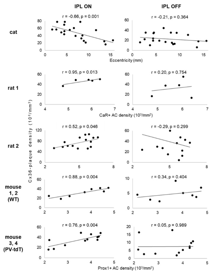
Figure 9.
Relationship of volumetric connexin-36 plaque density to retinal position in the ON- and OFF-sublaminae of the inner plexiform layer of mammalian retinas. Each scatterplot shows data from several locations within one or more retinas treated the same way. Retinal location is indicated by the distance from the area centralis for cat retina (top row). For rodent retinas, retinal location is indicated by the areal densities of amacrine cells labelled by CaR or Prox1. Higher cell density indicates a higher sampling density of the retinal image; these regions must therefore be functionally more “central”.

