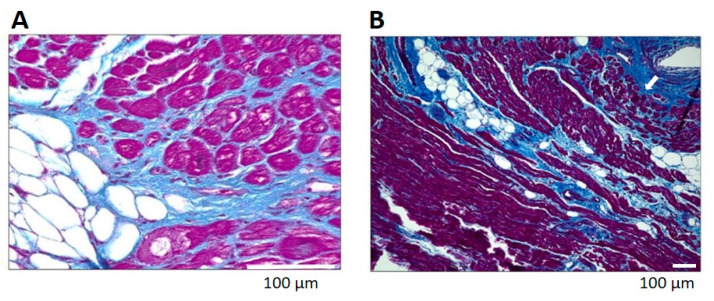
Figure 1.
Fibrosis in biopsies of left atria from patients with atrial fibrillation. Note pronounced perimysial and endomysial fibrosis (blue) in the subepicardial (Panel A) and perivascular (Panel B, arrow) myocardium. Masson’s Trichrome staining, adapted from Abe et al. [13].

