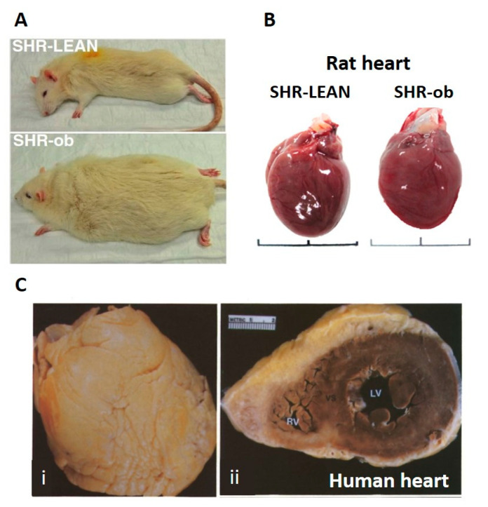
Figure 2.
Comparison of rat and human hearts, showing deposition of epicardial adipose tissue in human hearts, but not in rat hearts. Panel A: lean vs. obese (ob) spontaneously hypertensive rats (SHR), adapted from Linz et al. [68]. Panel B: hearts from lean vs. obese SHR. Note the absence of epicardial adipose tissue on each heart, regardless of animal weight. Adapted from Shiou et al. [69]. Panel C: heart of an 83-year-old woman (body–mass index 25.3; overweight) illustrating extensive epicardial adipose tissue covering the anterior surface (i). In cross-section (ii), the thickness of the adipose layer over the anterior aspect of heart is evident, while a thinner layer covers the posterior aspect of the heart. Adapted from Shirani et al. [70].

