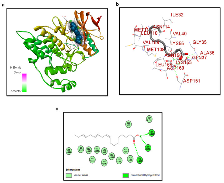
Figure 3.
Structure of p-JNK/ALA complex. (a) ALA docked in the binding site of JNK. α-helices and β-strands in the catalytic domain are shown in reddish and yellow, respectively. (b) shows the silver color ligand ALA and interactive residues. (c) Binding mode interaction of ALA with the active site residues. The green dotted lines are the conventional hydrogen bond of ASP169, ASN156, and LYS153 active site residues with ALA.

