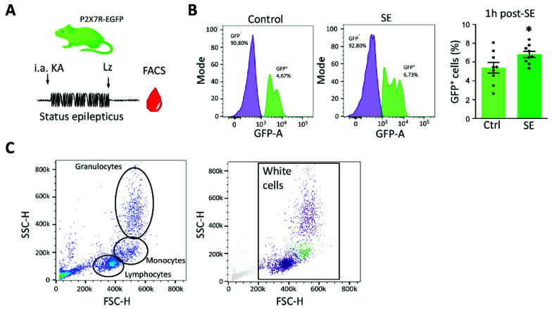
Figure 3.
Increased P2X7R expression in blood cells post-status epilepticus in mice. (A) Mice expressing P2X7 C-terminally fused to EGFP are subjected to intra-amygdala KA-induced status epilepticus. Blood was collected 1 h post-treatment with the anticonvulsant lorazepam. (B) Graph showing the histogram of the GFP-A (area) in control conditions and 1 h post-SE. The percentage of GFP+ cells is significantly increased in animals treated with intra-amygdala KA 1 h post-lorazepam (SE) when compared with vehicle-injected control mice (Ctrl) (N = 9 per group) (Unpaired Student’s t-test, p = 0.0493). (C) Forward side scatter indicating the distribution of the GFP+ cell population (in green). Note, size and complexity suggests the GFP+ cell population being composed mainly of monocytes. * p < 0.05.

