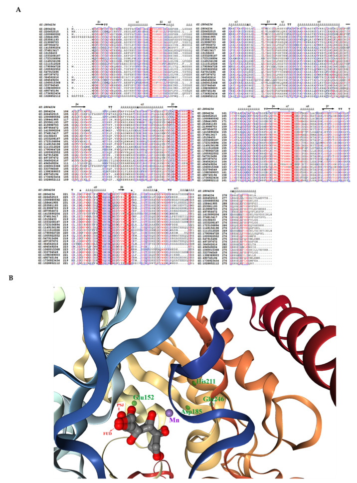
Figure 5.
(A) Multiple sequence alignment of d-tagatose-3 epimerases and d-allulose-3 epimerases. The red-boxed characters are the strictly identical amino acid residues. (B) Superimposition of the crystal structure of PDB:4PFH. The purple ball is the Mn2+ ion. The green dots are the ligand residues (in Chain B) with interactions with the Mn2+ ion and the substrate. The substrate d-fructose (FUD) and the product d-psicose (PSJ) are shown with arrows. The figure was visualized with an NGL engine powered by the MMTF program.

