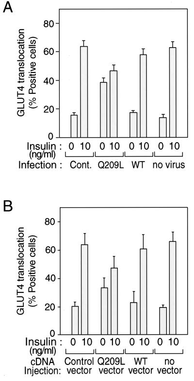
FIG. 3.
Effects of Gαq expression on insulin-induced GLUT4 translocation in 3T3-L1 adipocytes. Serum-starved 3T3-L1 adipocytes on coverslips were incubated with or without insulin (10 ng/ml) for 20 min after 60 h of Gαq-expressing adenovirus infection (MOI = 10) (A) or after 24 h of nuclear microinjection with Gαq expression vector and with GFP expression vector as a marker (B). Fixed cells were stained with rabbit anti-GLUT4 antibody and incubated with TRITC-conjugated anti-rabbit IgG antibody, as described in Materials and Methods. The percentage of cells positive for GLUT4 translocation was calculated by counting at least 100 cells at each point. The data are means and standard errors from three independent experiments. (A) Cont., mock control adenovirus; Q209L, Q209L-Gαq-expressing adenovirus; WT, wild-type-Gαq-expressing adenovirus. (B) Control vector, GFP-expressing vector only.

