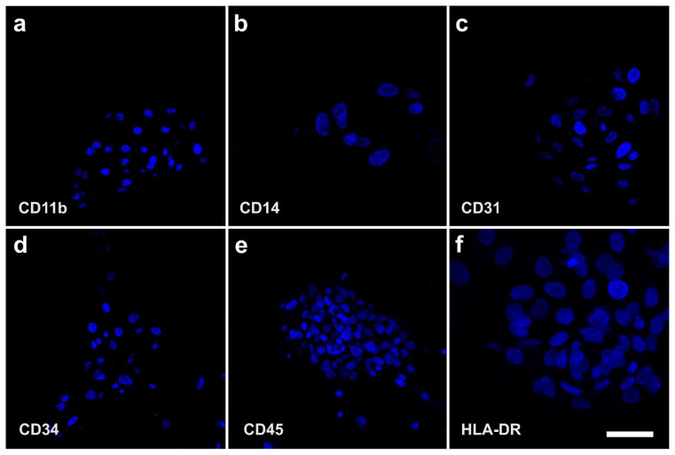
Figure 9.
Immunofluorescence detection of CD11b (a), CD14 (b), CD31 (c), CD34 (d), CD45 (e), and HLA-DR (f) on the surface of human adipose-derived stem cells that were cultured for four days in serum-free media (previously unpublished figure). The scale bar represents 60 µm in (a,d,e), 20 µm in (b), 40 µm in (c), and 30 µm in (f).

