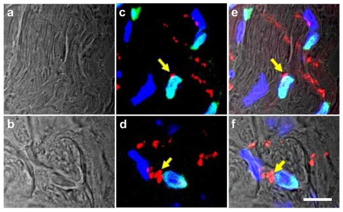
Figure 17.
Phase-contrast images (a,b), fluorescence images (c,d) and merged images (e,f) of heart sections prepared four weeks after experimental induction of myocardial infarction in severe combined immunodeficient (SCID) mice and injection of human adipose-derived stem cells (ADSCs) (a,c,e) or human adipose-derived regenerative cells (ADRCs) (b,d,f) into the peri-infarct region (adapted from [9] with permission from Oxford University Press). Cell nuclei were labeled with DAPI (blue); nuclei of human cells were labeled with an antibody against human lamin A/C (green); and gap junctions were labeled with an antibody against connexin 43 (red). Yellow arrows indicate detection of connexin 43 in gap junctions that could not be attributed to cell–cell contacts between mouse cells but most probably represented cell–cell contacts between mouse cardiomyocytes and descendants of injected human cells. The scale bar represents 15 µm in (a) and 10 µm in (b).

