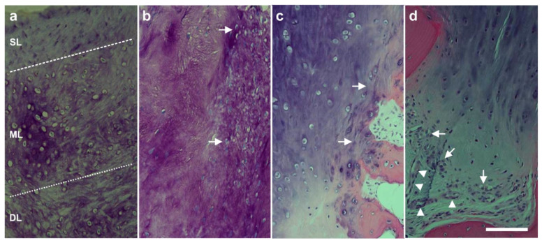
Figure 28.
Representative photomicrographs of 5-µm-thick histologic sections (stained with toluidin blue (a,b) or hematoxylin and eosin stain (c,d)) of the small tissue samples taken during arthroscopic inspection of the knees of the subject represented in Figure 26 at one year after arthroscopic removal of damaged cartilage and a single application of unmodified, autologous, adipose-derived regenerative cells (UA-ADRCs) (right knee) (a,c) or after performing a standard procedure without application of UA-ADRCs (i.e., arthroscopic removal of damaged cartilage and drilling of small holes into the bone) (left knee) (b,d), respectively (reprinted from [55] with permission from MDPI). The dotted lines in (a) (knee treated with UA-ADRCs) indicate a zonal organization of the newly formed cartilage with differently shaped chondrocytes in a superficial (SL), middle (ML) and deep layer (DL). The arrows in (b) (knee treated with a standard procedure without application of UA-ADRCs) point to scattered cells within newly formed amorphous fibrocartilage. The arrows in (c) (knee treated with UA-ADRCs) indicate typical chondrocytes with a small nucleus and a hollow space around in the directly built contact zone between the newly formed cartilage and bone. In contrast, the arrows in (d) (treatment without UA-ADRCs) point to an infiltration with inflammatory cells in the contact zone between a newly formed fibro-cartilage and bone. The arrowheads in (d) indicate three small blood vessels. The scale bar represents 100 µm in (a–d).

