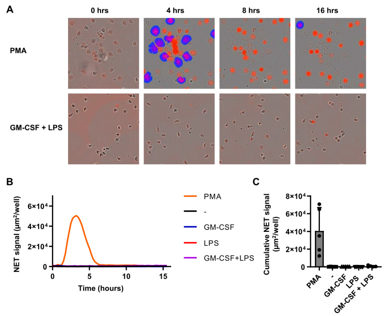
Figure 4.
Physiological stimuli do not induce NETosis. Neutrophils were cultured for 16 h in the presence of a cell impermeable fluorescent DNA-binding dye in the absence or presence of GM-CSF (50 U/mL) and/or LPS (10 ng/mL), or PMA (100 µg/mL). Fluorescence was measured every 15 min to determine NET formation and cell death. (A) Overlays of phase contrast and fluorescence images showing accessible DNA in red, and extracellular DNA (>400 µm2) in blue (NETs). Images are representative of 5 independent experiments. (B) NETosis was determined as fluorescence signal area in µm2 per well. Only areas larger than 400 µm2 were used for calculations (n = 5). (C) NETosis expressed as fluorescence area after 4 h of stimulation (n = 5). Data are presented as mean ± SD.

