Figure 5.
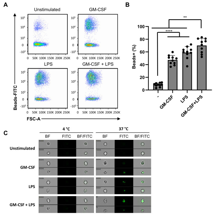
Phagocytosis is independent of dual stimulation. Neutrophils were stimulated for 2 h in the presence of FITC-labeled melamine beads and in the absence or presence of GM-CSF (50 U/mL), fMLF (1 µM), TNF (1 ng/mL), or LPS (10 ng/mL). (A) Phagocytosis was analyzed using flow cytometry. (B) Phagocytosis, determined by flow cytometry as percentages of FITC positive neutrophils (n = 10). (C) Imagestream microscopy images of neutrophils stimulated at 37 °C or 4 °C with GM-CSF (50 U/mL) and/or LPS (10 ng/mL) in the presence of FITC-labeled melamine beads. Location of phagocytosed beads is shown in green (left), and overlay of phase contrast neutrophil (right). Data are presented as mean ± SD. Asterisks indicate significant differences: ** p < 0.01 and **** p < 0.0001, one-way ANOVA with Tukey’s correction.

