Figure 6.
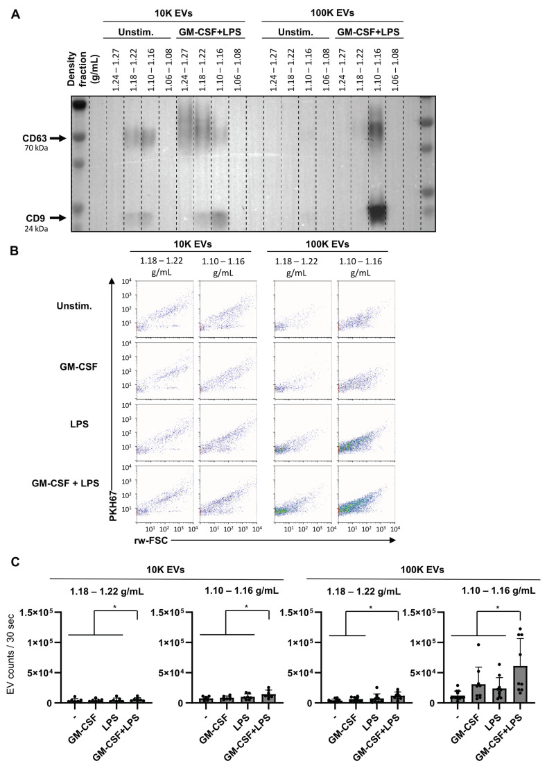
Dual stimulation of neutrophils increases EV release. Neutrophils were cultured for 2 h in the absence of presence of GM-CSF (50 U/mL) and/or LPS (10 ng/mL). After 2 h, culture supernatants were collected for the analysis of EV release. (A) Protein analysis (Western blotting) of EVs pelleted at 10 kg and 100 kg and floated into a sucrose gradient from neutrophils from a representative donor. Analysis is shown for CD9 and CD63 (tetraspanins; general EV-markers). (B) High-resolution flow cytometric analysis of purified PKH67-labeled EVs pelleted at 10 k and 100 k and floated in a sucrose density gradient from neutrophils stimulated with GM-CSF and/or LPS. (C) Quantification of EV release in the EV density fractions (1.10–1.16 g/mL and 1.18–1.22 g/mL) as determined by high-resolution flow cytometry. Indicated are the numbers of detected events within the fixed time frame of 30 s, with multiple conditions in each experiment. Data are presented as mean ± SD. Asterisks indicate significant differences: * p < 0.05, (n = 7−11), one-way ANOVA and mixed-effects analysis with Tukey’s correction.

