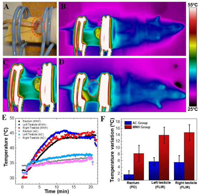
Figure 3.
(A) Positioning of the animal’s testicles on the magnetic field coil. Note: fiber-optic probes to measure testicular and rectal temperatures. (B) Thermographic image of an animal during the procedure of magnetic nanoparticle hyperthermia when the average temperature of the testicles was 45 °C. It is possible to visualize that the only heated part of the body is the testicular area. (C) Photograph of the testicular region of the same (B) MNH group animal. (D) Thermographic image of an AC group animal after 15 min of magnetic field exposure. Note that no heat generation was observed in the testicular region. (E) Temperature data of both animals, MNH group and AC group, during treatment. Rectal temperature was obtained using a fiber-optic thermometer, while the left and right testicle temperatures correspond to an average value obtained using the thermal camera. (F) Temperature increase in AC and MNH groups considering all animals investigated (mean ± SD).

