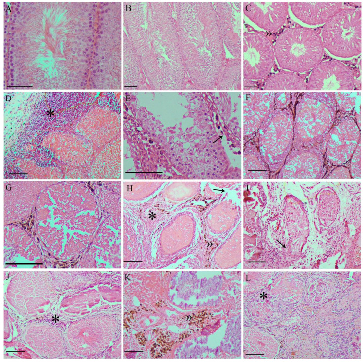
Figure 6.
Representative photomicrographs of testicles from the: Saline (A), AC (B), and MF (C) groups, presenting normal seminiferous tubules and all germline cell types. In the MNH-D7 group (D–F), the seminiferous tubules presented damage to the germinal epithelium and rupture of the basement membrane was observed (D), in addition to blood leaking into the tubular lumen and inflammatory infiltrate (*) in the interstitial tissue (E). It was possible to identify nanoparticle agglomerates in the interstitium (F). In the MNH-D28 group (G–I), seminiferous tubules showed coagulative necrosis (G), the interstitial connective tissue was thickened (H), and the typical tubular structure was lost (I). Nanoparticle agglomerates are still visible in the interstitium (G,H). In the MNH-D56 group (J–L), seminiferous tubules lost definition (J,K) and began to be replaced by connective tissue (L), the nanoparticle agglomerates are still visible (K). »: nanoparticle agglomerates; black arrows: seminiferous tubule basement membrane rupture; *: inflammatory infiltrate. Bars = 10 µm.

