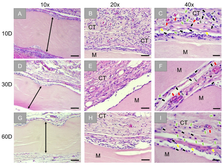
Figure 2.
Exemplary histological images of the subcutaneously implanted collagen-based sugar-crosslinked barrier membrane (Ossix® Plus) at three time points: 10 days (first row: (A–C)), 30 days (second row: (D–F)), and 60 days (third row: (G–I)). Stretched black arrow: compact layer of the membrane and M: membrane, CT: connective tissue, black arrows: macrophages, red arrows: blood vessels, green arrows: granulocytes, grey arrows: lymphocytes, yellow arrows: fibroblasts. (HE-stainings, 10×, 20×, and 40× objective magnifications with scale bars: 100 µm, 50 µm, and 20 µm, respectively).

