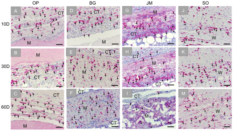
Figure 6.
Exemplary images of the immunohistochemical detection of anti-inflammatory M2 macrophages within the bed implants of the different barrier membranes and the sham operation group at three time points: 10 days, 30 days, and 60 days. First column: Ossix® Plus (OP) (A–C), second column: Bio-Gide® (BG) (D–F), third column: Jason® membrane (JM) (G–I), and forth column: sham operation (SO) (J–M). CT: connective tissue, M: membrane, W: wound area, black arrows: CD163-positive cells (CD163-immunostainings, 20× objective magnifications and scalebars = 50 µm).

