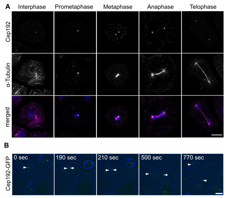
Figure 2.
Cep192 is present at the mitotic spindle poles. (A) Immunofluorescence microscopy of AX2 cells in interphase and indicated mitotic stages, stained with anti-Cep192 and anti-α-Tubulin. Secondary antibodies were anti-rabbit-AlexaFluor-488 and anti-rat-AlexaFluor-568, cells were fixed with methanol, DNA stained with DAPI. (B) Cep192-GFP is present during the splitting of the mitotic centrosome. Selected time points from Video S2 are displayed. Cells were viewed under agar overlay. Bars = 5 µm.

