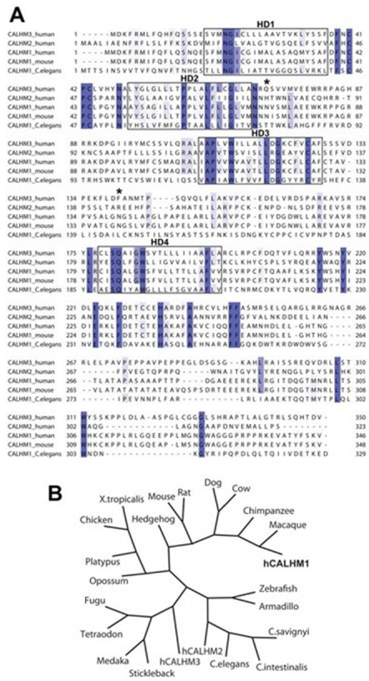
Figure 9.

The alignment and the phylogeny of the CALHM1. (A) the alignment of the sequence of the human CALHM3, CALHM2, and CALHM1, and of the murine and the C. elegans CALHM1. The conserved sequences have been highlighted with blue and the sequence conservation has been mapped with a gradient of color, the darkest color is used to represent the sequences having absolute level of identity and the lighter colors to represent the sequences having weaker level conservation. The boxes are to denote the hydrophobic domains 1–4 (HD1–4). Stars, the predicted sites of N-glycosylation on the human CALHM1. (B) the phylogenetic tree that include the human CALHM1, denoted as ‘hCALHM1′.
