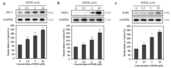
Figure 6.
BTDE increases the expression of HO-1, NQO1, and TrxR1. HaCaT cells were treated with BTDE (2.5–10 μM) for 24 h. The expression levels of HO-1 (a), NQO1 (b), and TrxR1 (c) were detected by Western blotting assay. Values are expressed as the mean ± SD of three independent experiments. ** p < 0.01, versus control group.

