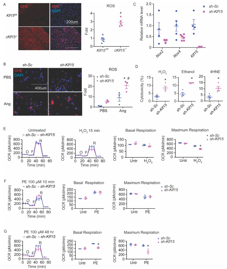
Figure 1.
KLF15 deficiency leads to increased susceptibility to ROS in the cardiomyocytes. (A) ROS in the hearts of cKlf15 KO and litter mate controls (8–10 weeks old, male) was measured by DHE staining (red) and DAPI (blue), and quantified using NIH Image J (n = 5, *: p < 0.0001 vs. litter mate controls). (B) NRVMs with shRNA knockdown of scrambled RNA (sh-Sc) or Klf15 (sh-Klf15). ROS was induced by treatment with Angiotensin II for 24 h and was measured by DHE staining (red) and DAPI (blue), and quantified using NIH Image J (n = 3, *: p < 0.01 vs. sh-Sc without treatment; #: p < 0.01 vs. sh-Klf15 without treatment). (C) qRT-PCR of Nox2, Nox4, and Klf15 in NRVM with sh-Sc or sh-Klf15 (n = 3–6). (D) NRVMs with shRNA knockdown of scrambled RNA (sh-Sc) or Klf15 (sh-K15) were subjected to different stimuli. Percentage of cytotoxicity derived from LDH assay after treatment with each stimulus is shown (ethanol: 100 mM 6 h; H2O2: 100 μM 2 h; 4HNE: 10 μM 8 h) (n = 4, *: p < 0.05 vs. sh-Sc). (E–G) Mitochondrial ETC function in NRVM assessed by Seahorse Mito Stress test using (E) H2O2 for 15 min, (F) phenylephrine (PE) for 10 min, (G) PE for 48 h, respectively (n = 3, *: p < 0.05 vs. sh-Sc, two-tailed Student’s t-test). The Holm–Sidak method was used to correct for multiple t-tests with alpha set as 0.05. Data are presented as mean ± SEM.

