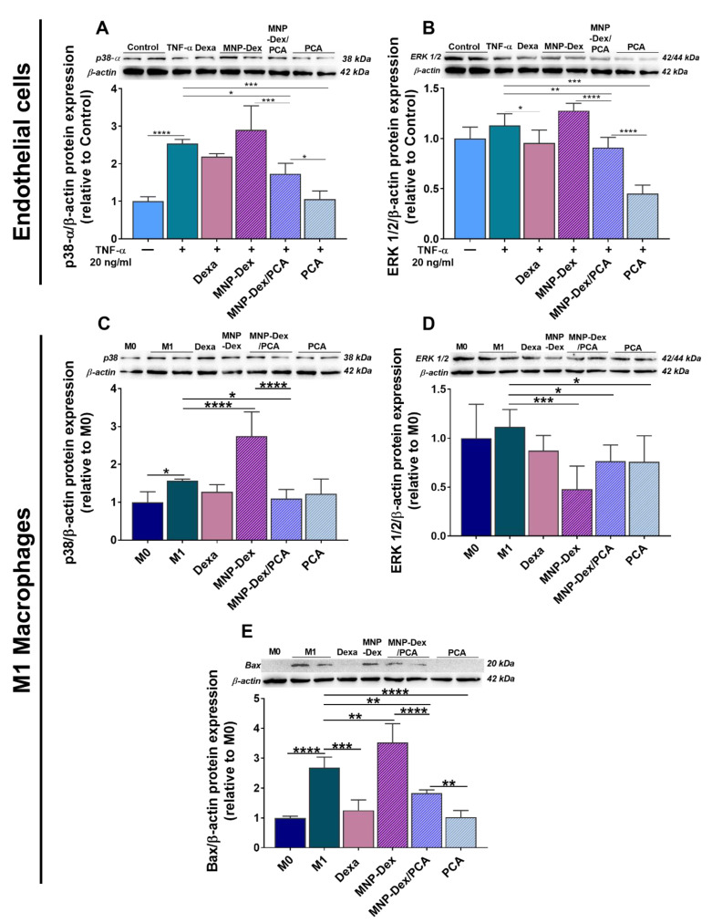
Figure 6.
Protein expression of p38-α (A,C) and total ERK 1/2 (B,D) in quiescent (‒) and TNF-α (20 ng/mL)-activated (+) EA.hy926 cells and M1 macrophages after 48 h of incubation with MNP-Dex/PCA (62 µg/mL MNP/ 350 µM PCA), as well as the protein expression of anti-apoptotic molecule Bax in M1 macrophages (E). Plain MNP-Dex, free PCA, and dexamethasone (Dexa) were used as controls. Protein expression was normalized to β-actin. The data are expressed as mean ± SD from three independent experiments. * p < 0.05, ** p < 0.01, *** p < 0.001, and **** p < 0.0001.

