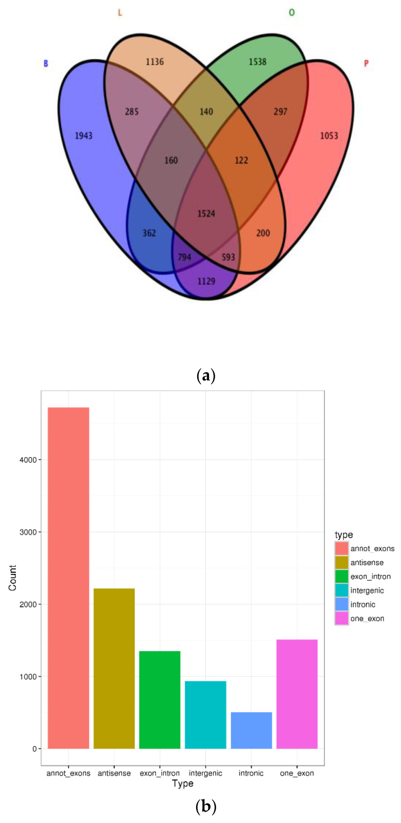
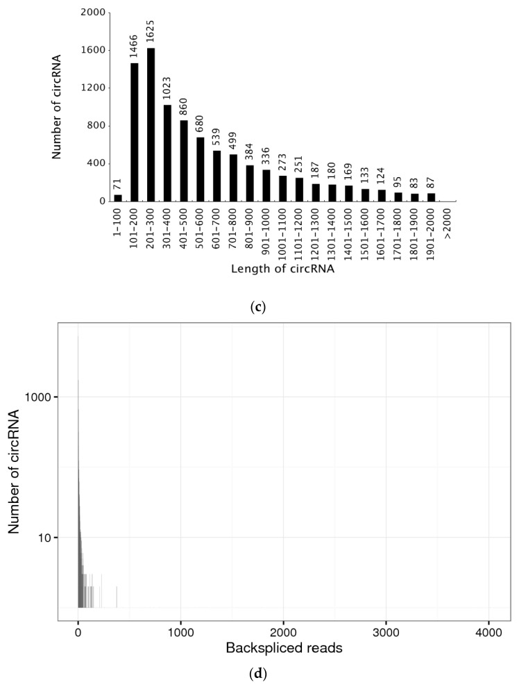
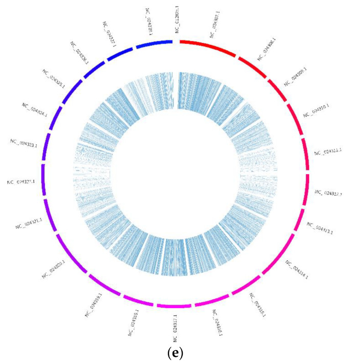
Figure 1.
Statistics of circRNA. (a) Venn diagram of circRNAs distributed in different tissues, brain (B), pituitary (P), liver (L) and ovary (O). (b) The type of circRNAs. (c) The length of total circRNAs. (d) The number of back-spliced reads of circRNAs. (e) Distribution of circRNAs in different chromosomes.


