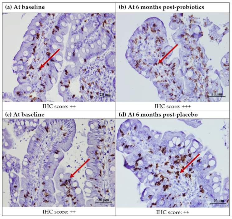
Figure 4.
Immunohistochemical staining of CD8+ protein in the duodenal mucosa (CD8+,400X). (a) Duodenal mucosa of a patient with NAFLD at baseline. (b) Duodenal mucosa of a patient with NAFLD after 6 months of probiotics. (c) Duodenal mucosa of a patient with NAFLD at baseline. (d) Duodenal mucosa of a patient with NAFLD after 6 months of the placebo. Red arrows show brownish CD8+ T lymphocytes in the lamina propria. Semi-quantitatively, no significant post-intervention difference was seen in either the probiotics of placebo groups. Staining score: 0: no staining; +: focal staining; ++: regional staining; and +++: no loss.

