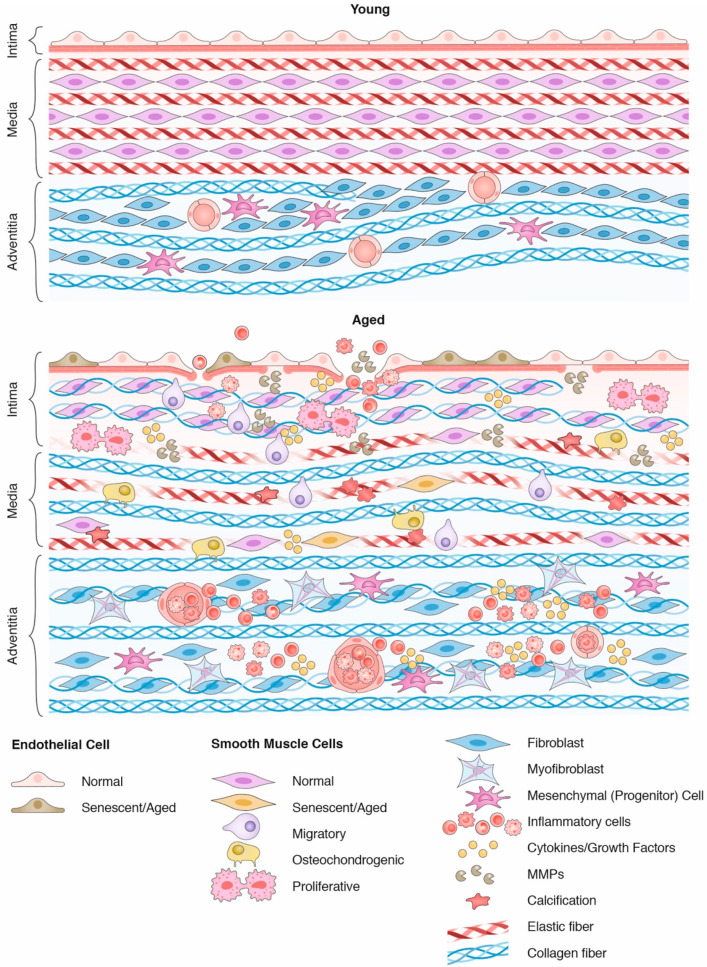
Figure 1.
Age-mediated changes in the arterial wall. The upper panel demonstrates the typical features of a young artery, formed by endothelial cells supported by a basement membrane (intima layer), followed by concentric SMC layers in association with elastic fibers, forming lamellar units in the medial layer. Fibroblasts and mesenchymal (progenitor) cells form the adventitial layer along with wavy collagen fibers and vasa vasorum. With aging (bottom panel), endothelial cells become senescent, facilitating the infiltration of inflammatory cells. In the medial layer, elastic fibers become calcified and fragmented, while collagen secretion increases, culminating in vascular stiffening. These changes expose SMC to greater mechanical stress, leading to a broad spectrum of phenotypes that only accentuate arterial aging through the senescence-associated secretory phenotype (SASP). In the adventitia, inflammatory cells use the vasa vasorum to infiltrate the arterial wall, while collagens distend and fibroblasts transdifferentiate into myofibroblasts.

