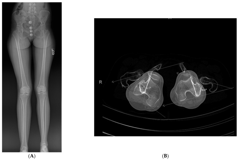
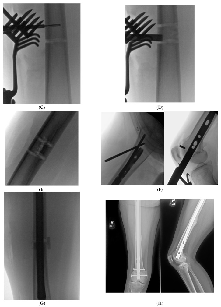
Figure 5.
(A): Full length standing radiograph. There is not a diaphyseal malunion; the patient has a 2 cm LLD, and rotational malalignment. The clamshell osteotomy was used to shorten and correct the rotational malalignment. (B): Computed tomography anteversion study demonstrating a 52° degree femoral anteversion and 30° femoral anteversion of the right and left lower extremity, respectively. (C): Intraoperative fluoro view showing bicortical drill holes being created in a diaphyseal segment measuring 2 cm. (D): An osteotome is used after the bicortical drill holes. Subsequently, a saw was used for the perpendicular cuts at the proximal and distal aspect of the osteotomy to create the clamshell. (E): Lateral fluoro view illustrating the clamshell osteotomy. No secondary fracture lines were propagated during this osteotomy. (F): 2.0 kirschner wire was placed in the proximal and distal aspect to be used as reference points when correcting the rotational malalignment. (G): AP fluoroscopic view after the clamshell segment was mobilized, and rotational malalignment was corrected. Her right knee was taken through range of motion after surgery to ensure there was not any patellofemoral maltracking. (H): AP and lateral femur XR demonstrating osseous healing of osteotomy at 6 months follow up.


