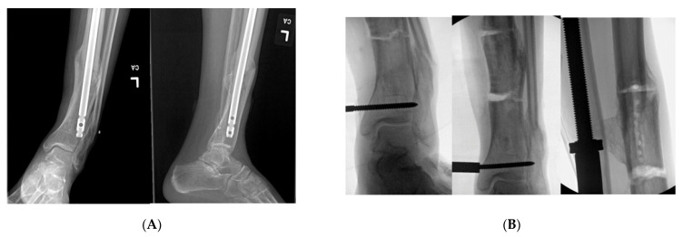
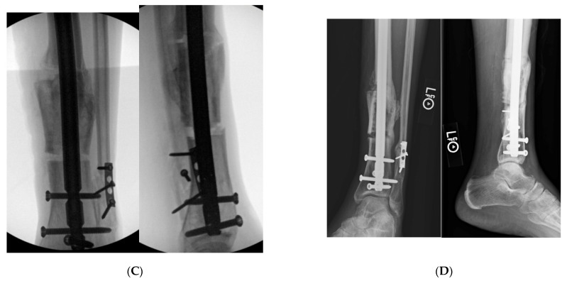
Figure 6.
(A): AP and lateral tibial XR demonstrating failure of tibial nail with valgus malunion. Notice there are not any distal interlocking screws. (B): Intraoperative fluoro views demonstrating medial universal distractor being used to assist with deformity correction, and maintain alignment during intramedullary nailing. (C): Intraoperative views demonstrating tibial nail and fibular plate after clamshell and fibular osteotomies. (D): AP and lateral 3-month post operative follow up XRs demonstrating healed clamshell osteotomy.


