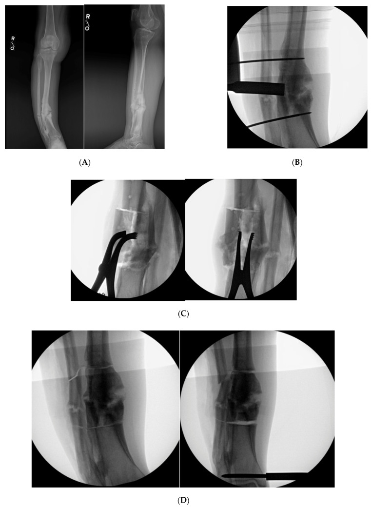
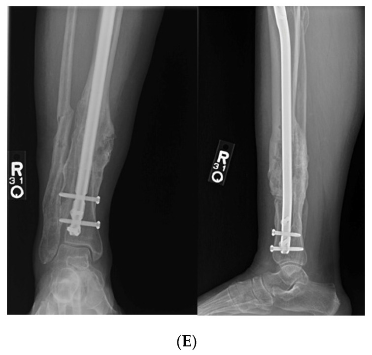
Figure 7.
(A): AP and lateral XR demonstration varus non-union deformity with segmental fibular fracture. (B): Intraoperative fluro view with threaded k-wire at proximal and distal aspect of malunited segment. A core reamer is being used to create the sequential bicortical drill holes. A core reamer can be used if the malunited segment is significantly larger than a 3.5 drill bit. (C): Note the lamina spreaders being utilized to open the osteotomized clamshell segment. (D): Medial universal distractor being utilized to assist with deformity correction. The distractor can be left in place during the nailing procedure. (E): AP and lateral 3-month postoperative radiographs demonstrating healed clamshell and fibular osteotomies.


