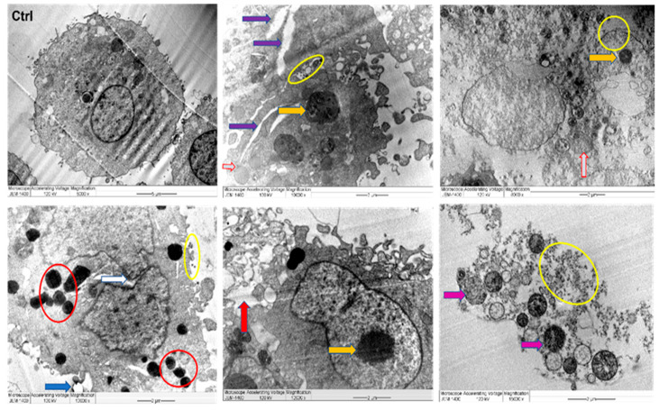
Figure 11.
TEM images of MDA MB 231 cell lines, where untreated cell (Ctrl) exhibiting rounded shape cell with complete organelles and nucleus. Image magnification ×6.000; bar 2 μm. Other images at various magnifications show the interaction of NPs and cancer cells leading to ultrastructural changes displaying early apoptosis characteristics such as chromatin condensation and nucleus cleavage (white arrow) and over whole-cell shrinkage as well as late apoptosis: lipid droplet (blue arrow), peroxisomes (red circle) and enlarged mitochondria (grey arrow), damaged mitochondria (violet arrow), and condensed nucleus (yellow arrow). In addition, damaged cancerous cells are observed with NPs located in the cytoplasm, outer cell, and nucleus membranes (yellow circle).

