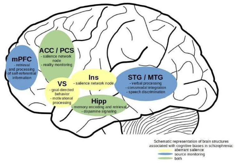
Figure 2.
Schematic representation of brain regions with effects detected between schizophrenia patients and non-clinical controls in fMRI studies of aberrant salience and source monitoring. For display purposes, effects are presented only on the left hemisphere of the brain. mPFC—medial prefrontal cortex, ACC/PCS—anterior cingulate cortex and paracingulate sulcus, vs.—central striatum, Ins—insula, Hipp—hippocampus, STG/MTG—superior and middle temporal gyri.

