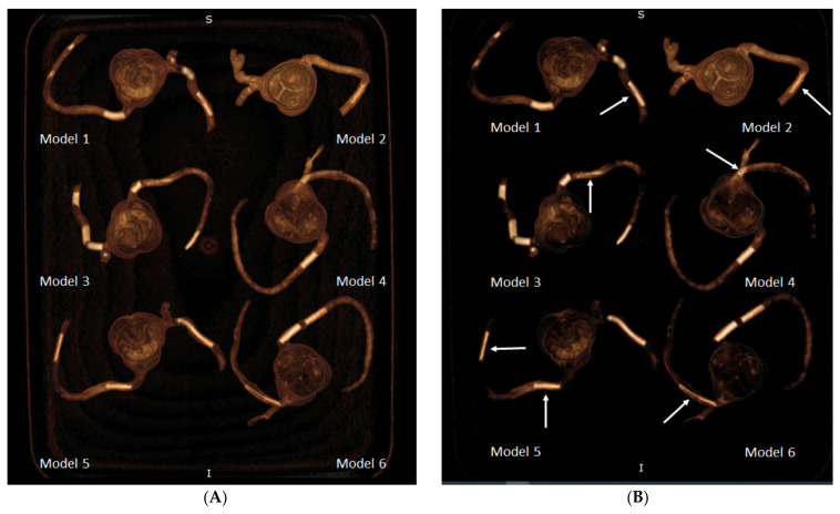
Figure 9.
3D volume rendering (VR) views showing calcified plaques in the coronary arteries. (A) VR views with wide window width (window width and window level: 1250 and 250) showing both plaques and coronary lumen in these 3D-printed models. (B) When narrow window width (window width and window level: 1050 and 210) is applied, high attenuation plaques are visualised more clearly, with some of the coronary lumen not displayed. This is especially apparent when visualising model 1 (plaque 2), model 2 (plaque 7), model 3 (plaque 13), model 4 (plaque 15), model 5 (plaques 18 and 19), and model 6 (plaque 20). Arrows refer to these plaques better visualised on VR images with narrow window width.

