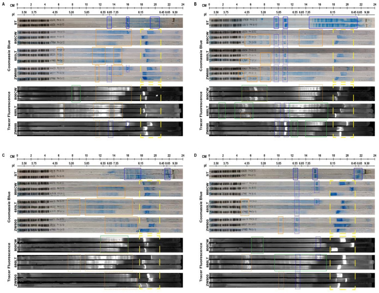
Figure 5.
Coomassie blue stain and fluorescent imaging results of completed immobilized pH gradient (IPG) strips. Unmodified reference (ST) and samples from each conjugate (800CW, 680LT, ZW800) were run on separate IPG strips. pI bands and patterns for each reference antibody are highlighted with a solid blue outline. In tracer strips with coomassie, bands that correspond to the reference antibody are given a dotted blue outline, while bands that are seen only after conjugation are given a solid orange outline. In tracer fluorescence, green solid outlines correspond to bands and patterns visible on the fluorescent scans but not on the coomassie-stained strips. Dotted blue and orange outlines correspond to bands marked before with solid blue and orange outlines in reference antibody or coomassie-stained conjugated samples, respectively. The dashed yellow outline highlights areas of the gel that are aspecifically stained as a result of antibody buffer components. Infliximab (B) and Ustekinumab (D) showed the best retention of their native bands, but all conjugations (vedolizumab (A), infliximab (B), adalimumab (C), ustekinumab (D)) resulted in shifts in primary band pI and formation of new bands. Infliximab showed much more acidic bands both natively and especially after conjugation to dyes. Extensive acidic shift may be related to instability in the conjugated protein. The scale of the gels is shown in cm and in pI. pI scale was based on a pI standard ladder (not shown) run on the same system alongside the samples. pI’s of sample bands are interpolated based on the position on the gel and the distribution of pI’s in the pI standard.

