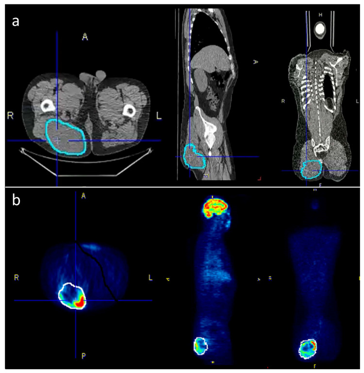
Figure 1.
To accurately outline the tumors, the VOIs were selected semi-automatically on the PET-images where possible and then manually adjusted to the tumor borders on the CT scan. (a) The corrected VOIs were then copied back to the PET-scan (b) in order to obtain identical VOIs for both PET and CT data.

