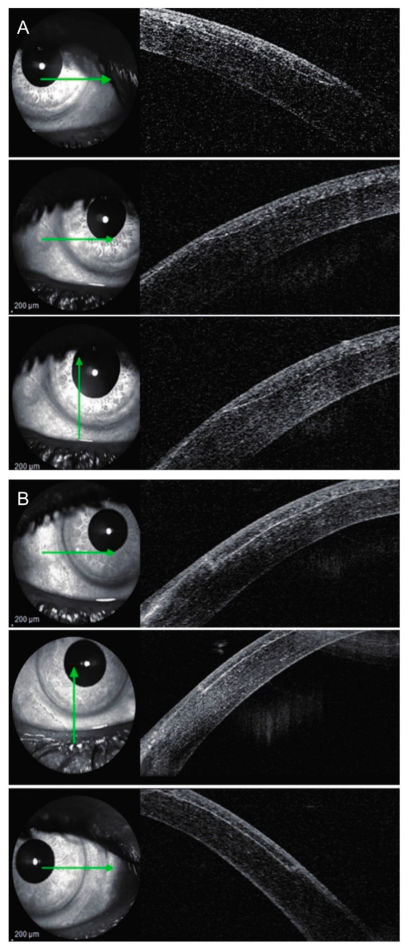
Figure 3.
Laser in situ keratomileusis (LASIK) flap side-cut angle and edge outline in (A) microkeratome-assisted LASIK and (B) femtosecond laser-assisted LASIK groups by anterior segment optical coherence tomography (OCT). Green arrows indicate the location from where the OCT scans were obtained. Reproduced with permission from [28].

