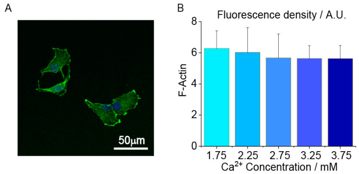
Figure 4.
The effects of extracellular Ca2+ concentration on human chondrocytes actin filament fluorescence density. (A) A representative chondrocytes actin fluorescence image taken at 1.75 mM Ca2+ concentration. Scale bar 50 μm; (B) No significant difference in the average fluorescence density of each group. 60 cells are measured and analyzed in each group. ANOVA and Tukey’s post hoc is performed for data analysis. Cell mechanics and cell–ECM adhesion are regulated by myosin and integrin expression at different [Ca2+]. Myosin, as one of the components of the cytoskeleton, can bond with actin and will influence the stiffness of cells (Figure 5A). During the migration, cells need to dissociate adhesions and form a new integrin–ECM adhesion (Figure 5B). The expression of myosin increased to the peak value from 1.75 mM to 2.75 mM, and then decreased from 2.75 mM to 3.75 mM [Ca2+] (Figure 5C). The expression of integrinβ1 increased in the lower [Ca2+] (e.g., 1.75 mM and 2.75 mM) and decreased in the higher [Ca2+] (e.g., 2.75 mM and 3.75 mM). (Figure 5D). The expression of integrinβ3 showed no difference between each group (Figure S2).

