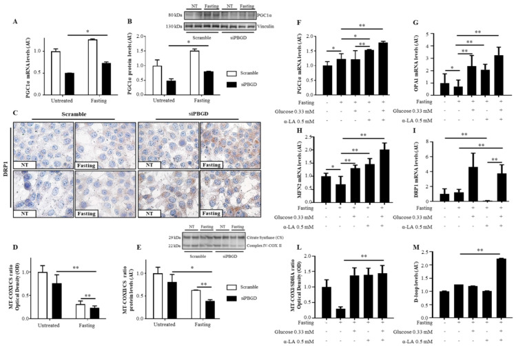
Figure 3.
α-LA combined to glucose recovered mitochondrial dynamics in PBGD-silenced HepG2 cells. (A,B) PGC1α mRNA and protein levels were assessed in both scramble and siPBGD cells at baseline and after fasting by qRT-PCR and Western blot, respectively. (C) Cytoplasmatic localization of DRP1 protein assessed at immunocytochemistry in scramble and siPBGD cells in the absence or in presence of fasting. (D) Mitochondrially-encoded subunit I (MT-COXI) of Complex IV was evaluated by ELISA (λ = 600 nm) and normalized to nuclear-encoded citrate synthase levels (λ = 405 nm). (E) Protein levels of mitochondrially-encoded subunit II (MT-COXII) of Complex IV was assessed by Western blot and normalized to nuclear-encoded citrate synthase. (F–I) PGC1α, OPA1, MFN2, and DRP1 expression was evaluated by qRT-PCR in siPBGD cells at baseline, after fasting, and in the presence of glucose, α-LA, or both treatments. (L) Intracellular MT-COXI of Complex IV was assessed by ELISA (λ = 600 nm) in siPBGD cells with or without fasting and pre-treated with glucose, α-LA, and α-LA + Gluc. MT-COXI protein expression was normalized to nuclear-encoded citrate synthase levels (λ = 405 nm). (M) D-loop levels were measured in DNA samples extracted from siPBGD cells at basal status and in fasting as well as in those treated with glucose, α-LA, and α-LA + Gluc. For gene expression, data were normalized to the ACTB housekeeping gene and expressed as fold increase (Arbitrary Unit—AU) compared to the control group. For Western blot, data were normalized on vinculin or citrate synthase housekeeping genes. At least three independent experiments were conducted. Adjusted * p < 0.05 and ** p < 0.01.

