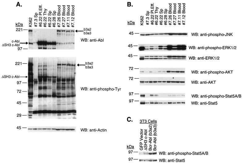
FIG. 7.
(A) Expression of Abl proteins and total tyrosine phosphorylation profiles in tumor cells. Lysates from equal numbers of cells, and 10 μg of lysate from K562 cells, were run on 6 to 15% polyacrylamide gradient gels, transferred to nitrocellulose filters, and probed with anti-Abl (Ab-3), antiphosphotyrosine (PY20), or antiactin (AC-40) antibodies, as indicated. Samples 7.26 and 7.27 [Bcr-Abl/p210 (b3a2)] and samples 7.11 and 7.12 [Bcr-Abl/p210 (b3a2)] are peripheral WBC lysates from mice that had developed a CML-like myeloproliferative disease. Pleural effusion (Pl.Eff.), spleen (Sp), and thymic lymphoma (Thy) lysate samples 6.22 are from a ΔSH3 c-Abl mouse that developed a T-cell leukemia and lymphoma. Spleen lysate 6.28 is from a ΔSH3 c-Abl mouse that developed a B-cell leukemia. Spleen lysate 7.3 is from a GFP vector mouse. (B) Activation of signaling pathways in tumor cells. The same lysates as probed in panel A were probed with anti-phospho-JNK (G-7), anti-phospho-p44/42 (Thr202/Tyr204) MAP kinases (Erk1 and Erk2), anti-p44/42 MAP kinases (Erk1 and Erk2), anti-phospho-Akt (Ser473), anti-Akt, anti-phospho-STAT5A/B (Y694/Y699) (8-5-2), and anti-STAT5 (G-2). (C) Activation of STAT5 in NIH 3T3 cells. Two days after infection with the indicated titer-matched virus, lysates were prepared from NIH 3T3 cells, run on 6 to 15% polyacrylamide gradient gels, transferred to nitrocellulose filters, and probed with anti-phospho-STAT5A/B (Y694/Y699) (8-5-2), and anti-STAT5 (G-2). WB, Western blot.

