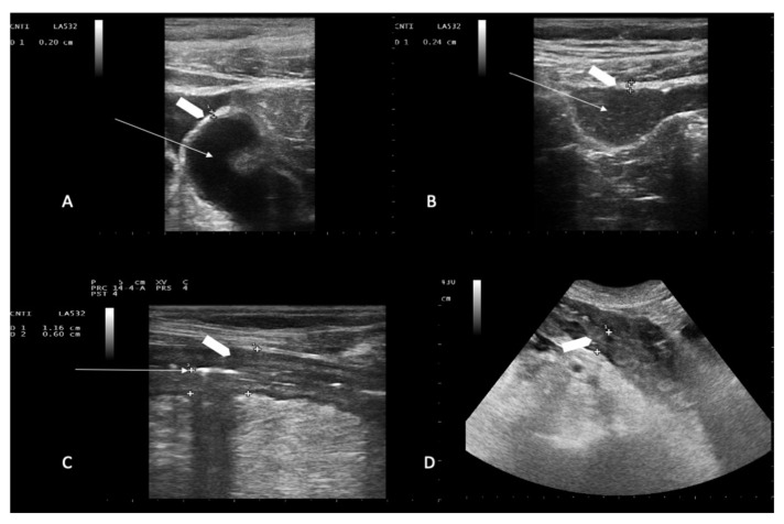
Figure 1.
Representative ultrasound imaging of ileum loops. Chemotherapy-induced diarrhea: ileum loops filled with liquid (panel A) and mixed solid/liquid material (panel B). White arrows indicate the lumen; white arrowhead indicates normal bowel wall thickness (2.0 mm in panel A, and 2.4 mm in panel B). Neutropenic enterocolitis involving the terminal ileum loop and descending colon: white arrow in C shows compressed lumen (panel C); white arrowheads show bowel wall thickness (6.0 mm in panel C, and 13 mm in panel D). Bowel wall layers are recognizable in (panel C) while boundaries are poorly defined in (panel D).

