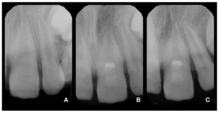
Figure 3.
X-rays of tooth 21. (A)—diagnostic X-ray of tooth 21; (B)—postoperative X-ray of tooth 21; (C)—control X-ray at 24-month follow-up. The continuing development of the root and narrowing of the root canal is visible. In addition, the formation of a radiopaque bridge under the calcium silicate cement and the absence of periapical radiolucency are apparent.

