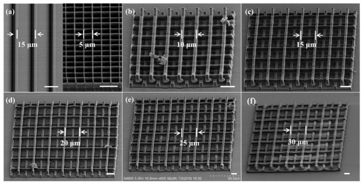
Figure 4.
SEM images of different microstructures. (a) Hydrogel line array (left) and ST scaffold (right). (b–f) A series of the 3D microscaffolds with different strut spacings of 10 μm, 15 μm, 20 μm, 25 μm, and 30 μm, with corresponding porosities of 69.7%, 79.5%, 84.4%, 87.4% and 89.3%, respectively. The scale bar is 10 μm.

