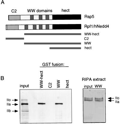
FIG. 7.
(A) Schematic representation of yeast Rsp5 and human Rpf1/Nedd4. GST-Rpf1 fusions to the regions of Rpf1 indicated by the solid bars were made. (B) (Left) HeLa cell extract was prepared in NP-40 lysis buffer (see Materials and Methods). The binding of hRpb1 to GST-Rpf1 fusion proteins immobilized on glutathione-Sepharose was analyzed by SDS-PAGE and immunoblotting. The “input” shows hRpb1 in the extract with forms IIo, IIa, and IIb. (Right) Similar experiment, with HeLa cell extract prepared in radioimmunoprecipitation assay (RIPA) buffer. The input and binding to GST-WW are shown.

