Figure 3.
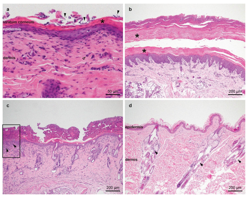
Histopathological images of representative skin biopsies, hematoxillin and eosin stain. (a) The epidermis is mildly hyperplastic and covered by compact parakeratotic keratin, characterized by the presence of retained nuclei in the stratum corneum (asterisk). There are numerous yeasts in between the corneocytes, indicated by the filled arrows. There is a mild perivascular infiltrate in the superficial dermis composed mostly of mast cells (empty arrow). (b) The epidermis of the paw pad is mildly hyperplastic and covered by a thick layer of compact parakeratotic keratin (asterisks). (c) The epidermis is mildly hyperplastic and covered by a thin layer of compact parakeratotic keratin. The stratum corneum is covered by a thick serocellular crust, composed of corneocytes, proteinaceous material, degenerate inflammatory cells, and abundant cocci. In the rectangle on the left edge of the image, an ulcer is present, characterized by the lack of the epidermis and abundant inflammatory cells in the superficial dermis underneath the ulcer (filled arrows). (d) Skin of a healthy cat as control showing the epidermis of normal thickness covered by basketweave orthokeratotic keratin together with the dermis and the adnexa (filled arrows).

