Figure 1.
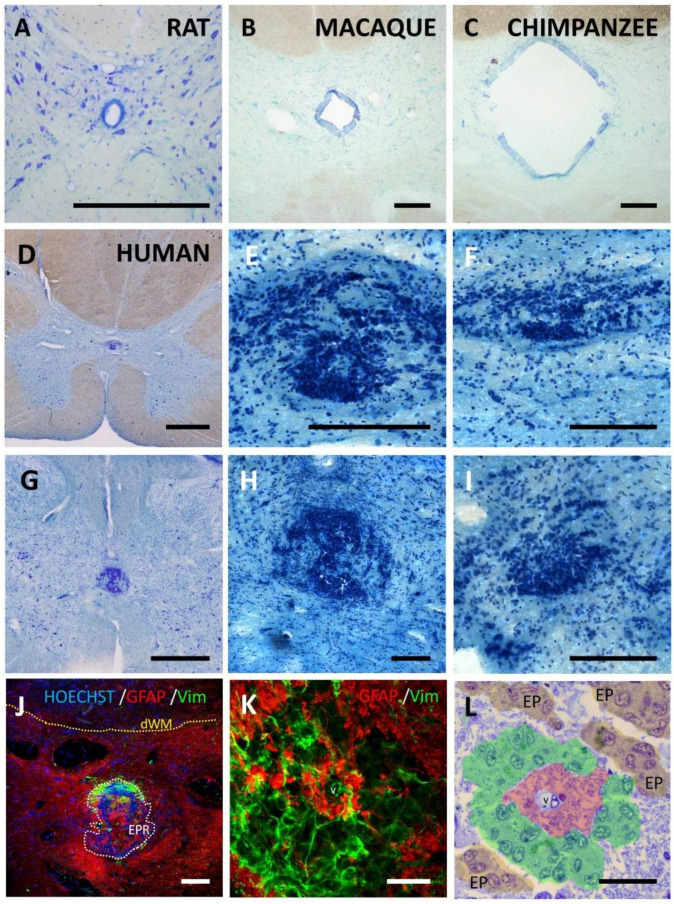
Ependymal region organization in different mammal species. In toluidine blue-stained sections, ependymal lining surrounding a patent central canal can be observed in most mammals such as rat (A), macaque (B) or chimpanzee (C). (D) Young and adult humans, on the contrary, mostly show absence of central canal which is substituted by a new organization of the ependymal region that includes large accumulation of cells (D–I). Examples from different individuals are shown: (D) male, 52 years (A10/017 sample); (E,F) higher magnification details of different spinal levels of (D); (G) male 39 years (A10/044 sample); (H) higher magnification detail of G; (I) Male 47 years (A10/067 sample). (J) In adult humans, the new structure in the ependymal region (EPR) substituting former canal also includes strong astrogliosis (GFAP immunoreactivity, red) and the presence of (K,L) perivascular pseudorosettes, i.e., cells expressing vimentin (green) radially oriented around a central vessel (v), separated from it by a hypocellular GFAP+ region. Pseudocolors in L highlight the GFAP hypocellular region (red) that surrounds the vessel (v), cells radially oriented (green) and ependymal cell groups around the perivascular pseudorosette (EP). dWM, dorsal white matter; Vim, vimentin. Magnification bars: (A), 250 µm; (B,C,E,F,H–J), 200 µm; (D,G), 1 mm; (K,L), 25 µm.

