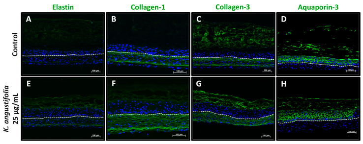
Figure 5.
Aging markers visualized by immunofluorescence staining. Elastin (A,E), collagen-1 (B,F), collagen-3 (C,G) and aquaporin-3 (D,H) expression, which usually decreases during skin aging, is shown in healthy skin substitutes untreated (A–D) and treated with the K. angustifolia extract at 25 μg/mL (E–H). The nuclei were stained with DAPI (blue). The dotted line represents the separation between the epidermis and dermis. Two substitutes for each condition were analyzed and confirmed with three different cell populations (N = 3, n = 2; scale bar: 100 μm).

