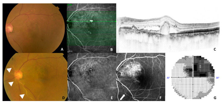
Figure 5.
A representative case (77-year-old woman) showing an adverse event during the follow-up. (A–C) At baseline, fundus photography showed an orange-red lesion superior to the fovea (left), a horizontal optical coherence tomography (OCT) scan demonstrated a double layer sign and retinal pigment epithelial protrusion (left) corresponding to the branching vascular network and a large polypoidal lesion on indocyanine angiography (ICGA) (middle). Whitened retinal vessels as indicated triangles were seen on color fundus photography (D) and a residual polypoidal lesion was seen on ICGA. (E) Partial perfusion as indicated a white arrow was seen in the inferior arteriole arising from the optic disc on fluorescein angiography 1 month after the third brolucizumab injection. (F) Almost half of the superior visual field defects were detected on the Humphrey visual field analyzer 30-2 program. (G) BCVA in the left eye was maintained at 0.7, in the decimal format, as was the baseline BCVA.

