Figure 1.
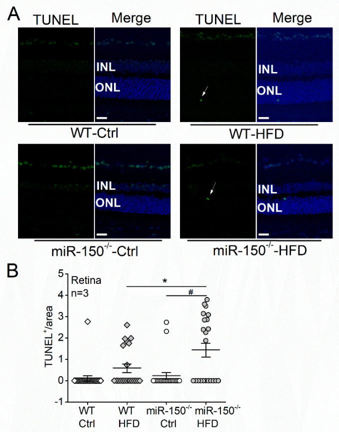
MicroRNA-150 knockout (miR-150−/−) exacerbates apoptosis in the diabetic retina. (A) Wild-type (WT) and miR-150−/− mice were fed on normal chow (Ctrl) or high-fat diet (HFD) for 6 months. Terminal deoxynucleotidyl transferase dUTP nick end labeling (TUNEL) staining of mouse retinal sections shows TUNEL positive (TUNEL+) apoptotic cells in green fluorescence. The arrows indicate apoptotic photoreceptors in the outer nuclear layer (ONL). The 4′,6′-diamidino-2-phenylindole (DAPI; blue) stains the cell nuclei. Scale bar: 20 µm. (B) The number of TUNEL+ photoreceptors was counted from 5–10 regions for each retinal section and normalized to the ONL area. WT-Ctrl: open square; WT-HFD: gray square; miR-150−/−-Ctrl: open circle; miR-150−/−-HFD: gray circle. * and # indicate statistical significances specified with a horizontal line. Each group has n = 3 (mice). p < 0.05, one-way ANOVA.

