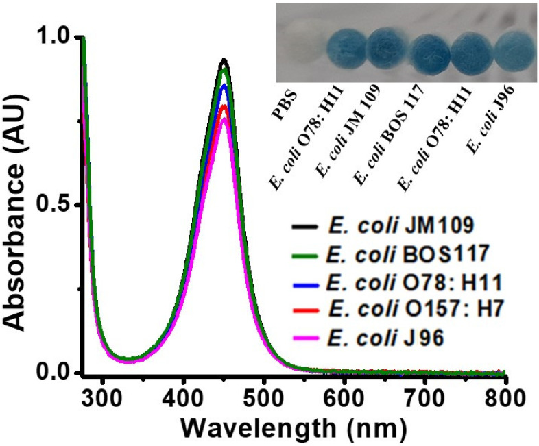
Figure 4.
UV–Vis absorption spectra of the samples (0.2 mL) containing different E. coli strains (OD600 of ~1), obtained after reaction with H2O2 (11.2 mM) in the presence of TMB (1.25 mM) at pH 3, followed by the addition of sulfuric acid (2 M, 2 µL) to stop the reaction. (Inset) shows the photograph of the cotton swabs containing E. coli strains (OD600 of ~1) obtained after reaction with H2O2 (11.2 mM) in the presence of TMB (1.25 mM) at pH 3. The reaction was conducted at room temperature (~25 °C).

