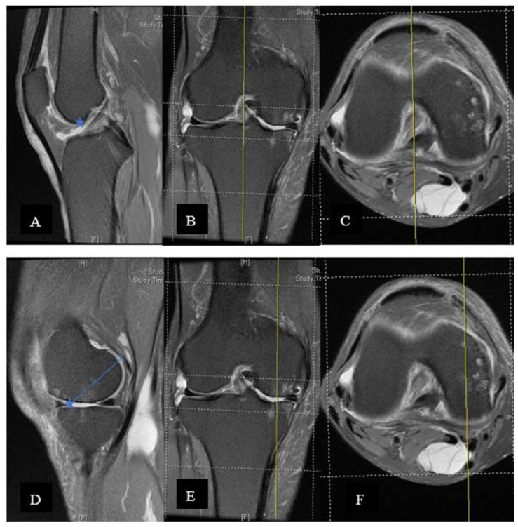
Figure 5.
(A–C) On the sagittal section, the cut that bisected the trochlea was obtained, confirming the axial and coronal sections. The corner between trochlea and blummensant line was identified and marked (star). (A) Central sagittal cut of the knee MRI. (B) Coronal cut of the knee MRI. (C)Axial cut of the knee MRI. (D–F) The marked point was transferred to the sagittal section at the central cut of the medial femoral condyle (also have to check on the axial and coronal planes). The marked point would estimate the most anterior point of the medial femoral condyle. The most posterior point included the visualised and marked cartilage. A full thickness loss of cartilage is usually observed at the anterior region. The cartilage thickness should be added back. (D) Sagittal cut through medial femoral condyle of the knee MRI (E) Coronal cut of the knee MRI (F) Axial cut of the knee MRI.

