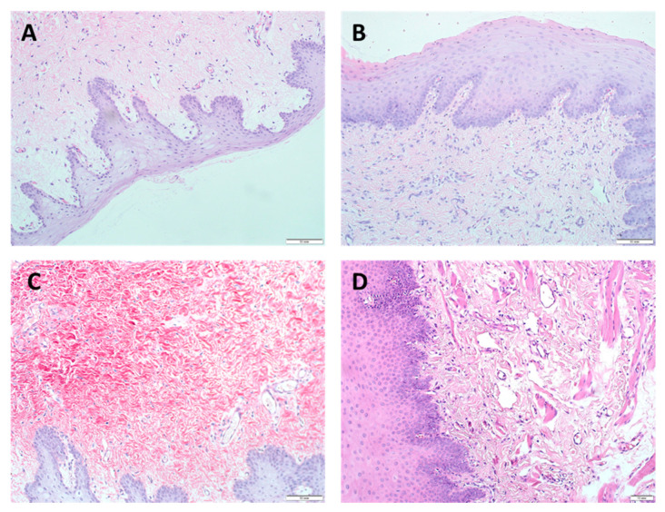
Figure 3.
Morphological characteristics of regenerative changes on the 7th day after augmentation of the gingival mucosa with Mucoderm material, H&E staining (A–C) magn. ×200, scale bar 50 μm; (D) magn. ×100, scale bar 10 μm: (A) surface augmentation without local injection of MSCs (SDW subgroup), intact integumentary squamous epithelium without signs of regenerative changes, angiogenesis in the form of separate capillary vessels, weak lymphohistiocytic infiltration, and fibroblastic responses were observed; (B) surface augmentation in combination with local injection of MSCs (SDI subgroup), signs of regenerative changes in the epithelium, increased angiogenesis, lymphohistiocytic infiltration, and fibrogenesis in the absence of changes in the fibroblastic response were observed; (C) under the flap without local injection of MSCs (UDW subgroup), morphological changes did not differ from those observed in the SDW subgroup implanted with the use of only Mucoderm by the surface augmentation; (D) under the flap in combination with local injection of MSCs (UDI subgroup), increased hyperplasia with foci of hyperkeratosis and acanthosis of the integumentary squamous epithelium, significantly higher angiogenesis, a decrease in lymphohistiocytic infiltration, and no changes in the fibroblastic responses were observed. A feature of the tissue reaction was the appearance of myofibroblastic foci in the submucosal layer, indicating a high regenerative activity of the tissue, possibly with the participation of MSCs.

