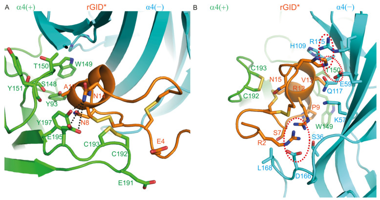
Figure 5.
Molecular model of the complex between rGID* and the α4(+)α4(−) interface. (A) Interactions between the receptor and Glu-4 (E4), Asn-8 (N8), Ala-10 (A10) and Asn-14 (N14) of rGID*; (B) Interactions between the receptor and Arg-2 (R2), Ser-7 (S7), pro-9 (P9), Val-13 (V13), Asn-15 (N15) and His-17 (H17) of rGID*. Hydrogen bonds are displayed as dotted black lines on the structure. Interactions between charged side chains are circled with a dotted red line.

